Saturday, April 21, 2007

Biotinidase Deficiency

Biotinidase deficiency is an inherited disorder in which the body is not able to process the vitamin biotin properly. Biotin is an important water-soluble vitamin that aids in the metabolism of fats, carbohydrates and proteins. 1 in every 110, 000 babies will have the deficiency.
Newborns suffering from biotinidase deficiency will appear normal at birth. The deficiency develops over time ranging from a few weeks to several years of age.
Biotinidase deficiency is an autosomal recessive disorder affecting both males and females. In individuals with this disorder, both copies of the biotinidase gene are defective. Both parents of an affected child have one abnormal copy of the gene, but usually do not show symptoms because they also have one normal copy. The normal copy provides approximately 50% of the usual enzyme activity, a level adequate for the body's needs. Individuals with one abnormal copy of the gene and 50% enzyme activity are said to be carriers or heterozygotes. As is typical of autosomal recessive inheritance, their risk for having another child with the disorder is 25% in each subsequent pregnancy.

Biotin deficiency can result in behavioural disorders, lack of coordination, learning disabilities, hearing loss, developmental delay and seizure.

Without biotinidase activity, biotin cannot be separated from foods and hence cannot be used by the body. Biotinidase enzyme also recycles biotin from enzymes that are important in metabolism.
HOWEVER, there is no need for much worry as it can be treated with oral biotin supplementation, preventing development of critical symptoms.

Here is the chemical structure of biotin:

Iodine Deficiency

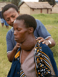
Iodine is an essential trace element (a chemical element that is needed in small quantities for proper growth, development and physiology aka micronutrient). When there is little iodine in our diet, iodine deficiency gives rise to goiter (swelling in the neck - below) and cretinism (severely stunted physical and mental growth) resulting in developmental delays and health problems.

Mild iodine deficiency has been reported to reduce intelligence quotients (I.Q.) by 10%-15% and cause increased rates of stillbirths and infant mortality


So where does the iodine in our diet come from?
Although the element is quite rare, certain plants have the ability to concentrate it from seawater, thus introducing iodine into the food chain.

Iodine is widely available in:
  • Cod
  • Sea bass
  • Haddock
  • Perch
  • Kelp
  • Dairy products
  • Plants grown in iodine-rich soil

In areas where little or no marine food is eaten, the people will suffer from iodine deficiency. To combat this, iodine deficiency is combatted by the additions of iodine to small amounts of table salt, known as iodized salt, and other foodstuffs such as flour.


Did you know?
Approximately two billion people are affected by iodine deficiency in this world, particularly in India, with 500 million suffering from deficiency.

Here are the recommended intakes for iodine:
  • Infants - 40 to 50 micrograms
  • Children (1 to 3 YO) - 60 micrograms
  • Children (4 to 6 YO) - 90 micrograms
  • Children (7 to 10 YO) - 120 micrograms
  • Children (11 years and above) - 150 micrograms
  • Pregnant women - 175 micrograms
  • Adult men and women - 100 to 200 micrograms

Monday, April 16, 2007

Primary Immune Deficiency Disorder

A Primary Immune Deficiency Disorder is present when a body part is missing or is unable to function normally, these are caused by genetic and intermal problems, increasing susceptibility to infections. It is a broad heading for many World Health Organisation recognised deficiencies.
It has been estimated that there are 100 different primary immunodeficiency diseases. All are genetic conditions in which specific cells of the immune system do not function properly. Clinical symptoms range from mild or nonexistent as in the case of selective IgA deficiency to severe symptoms as in the case of severe combined immunodeficiency (SCID), commonly referred to as "bubble-boy" syndrome.
Possible types of Primary Immune Deficiency Disorder:
Antibody Deficiencies An antibody is a protein made by white blood cells called B lymphocytes and plasma cells. Its function is to recognize and mark foreign microorganisms. It can be diagnosed with frequent and severe infections caused by organisms that do not affect people with healthy immune systems.
Complement Deficiencies The complement system consists of a group of proteins that attach to bacteria and viruses.
These proteins work together to disable and kill invading pathogens. People deficient in one or another of these proteins have a diminished ability to kill invaders, resulting in increased infections. Such people may also develop antibodies that react against the body's own cells and tissues, resulting in autoimmune diseases with immune-mediated damage to the body. The most common of these deficiencies is:
C2 Deficiency-an inherited defect in the gene for the complement protein called C2. This defect can cause an autoimmune disease such as systemic lupus erythematosus or can result in severe infections such as meningitis. The illnesses usually appear in childhood or in early adulthood.
Phagocytic Cell Deficiencies Phagocytes: white blood cells called neutrophils and macrophages that engulf and kill microorganisms. If defective either in their ability to kill pathogens or in their ability to move to the site of an infection. An increase in infections can happen. The most severe form is:
Chronic Granulomatous Disease-inherited deficiencies of molecules needed by neutrophils to kill certain infectious organisms. These illnesses usually appear early in childhood but may appear as late as adolescence. People with chronic granulomatous disease develop frequent and severe infections of the skin, lungs, and bones and develop localized, swollen collections of inflamed tissue called granulomas.
Due to healthcare advances, most patients with a Primary Immune Deficiency Disorder will be able to live normal lives to adulthood; however, they will have to rely on life-long treatment, such as intravenous gamma globulin infusions, antibiotic therapies, or bone marrow transplantation. Although the susceptibility to infections is a major consequence of the primary immunodeficiency diseases, they may cause other health problems as well, including allergies, asthma, swollen joints, digestive tract problems, growth problems or an enlarged liver and spleen.
The primary immune deficiency disorders range in frequency. Some disorders such as selective IgA deficiency are quite common, occurring as often as 1/500 to 1/1000 individuals. Others disorders such as SCID are rare affecting perhaps one person per million. Approximately 25,000 to 50,000 Americans are severely affected by primary immune deficiency disorders.
For more information:
http://www.primaryimmune.org/pid/1996_who.pdf -a very difficult report from the WHO.
Source:
http://www.primaryimmune.org/pid/whatis_pid.htm
http://www.medhelp.org/NIHlib/GF-739.html
http://www.medterms.com/script/main/art.asp?articlekey=25189
http://www.aaaai.org/patients/topicofthemonth/0407/

Sunday, April 15, 2007

Arginase Deficiency

Arginase deficiency is thought to be the least common of the urea cycle disorders. Two separate isozymes of the enzyme arginase exist. Type I is found in the liver and contributes the vast majority of hepatic arginase activity, while type II is inducible and found in extrahepatic tissues. The disease is caused by deficiency of arginase type I in the liver.

The hepatic urea cycle is the major route for waste nitrogen disposal, which is generated chiefly from protein and amino acid metabolism. Low-level synthesis of certain cycle intermediates in extrahepatic tissues makes a small contribution to waste nitrogen disposal as well. A portion of the cycle takes place in mitochondria. Overall, activity of the cycle is regulated by the rate of synthesis of N-acetylglutamate, the enzyme activator that initiates incorporation of ammonia into the cycle.

The reaction normally mediated by arginase is the terminal step in the urea cycle, which liberates urea with regeneration of ornithine. Consequently, as in argininosuccinic aciduria, both waste nitrogen molecules normally eliminated by the urea cycle are incorporated into the arginine substrate molecule in the reaction.

Think that is complicated? Look at THIS.

Photo Sharing and Video Hosting at Photobucket

As an inherited disorder, the age of onset is typically during the neonatal period(or in simpler terms, the early part of infancy). Because of its atypical form of manifestation, the disease may be easily missed in the neonatal period and only recognized in later infancy or early childhood. Some cases likely go undiagnosed, with clinical symptomatology attributed to cerebral palsy. Also, being an inherited disorder, parents have 1 in 4 chances of having an affected child with each conception. Affected infants and children may exhibit mental retardation, seizures, and spasticity (difficulty in walking).Luckily, arginase deficiency hardly results in death. yay!

Diagnosis is made by special blood tests called plasma amino acids. Enzyme level can be measured in the blood however specialized laboratory help is required for this. In some situations DNA mutation analysis is also available. To prevent giving birth to an affected child, prenatal diagnosis is possible based on DNA mutation analysis.

But if YOU have arginase deficiency (touch wood), this is what you should do!
1. Normalize plasma arginine levels
2· Go for genetic counselling
3· Take a low protein diet
4· Take these drugs: Sodium phenyl butyrate/ benzoate to divert nitrogen from urea cycle

Sunday, April 1, 2007

Introduction

A deficiency disease is basically a disease, such as rickets or scurvy, that is caused by a dietary deficiency of specific nutrients, especially a vitamin or mineral. The disease may stem from insufficient intake, digestion, absorption, or utilization of a nutrient.

In our following posts, we shall elaborate on several defiency diseases known to man.