There are two main forms of beriberi, which is caused by lack of Vitamin B1. When there is a low calorie intake and high levels of inactivity beriberi tends to affect the central nervous system.
This is known as dry beriberi and can eventually lead to paralysis. Less serious cases result in poor coordination and the tightening of muscles.
The second form, known as wet beriberi, is linked with very high activity levels and eating large amounts of carbohydrates. Symptoms include sweating, high heart rates and warm skin which if not treated lead to heart failure and death. Extremely high levels of B1 deficiency can lead to less blood flow into the brain, a condition called Wernicke-Korsakoff syndrome. This can cause confusion and involuntary movements of the eyes, which if untreated leads to coma and eventual death.
Beriberi is uncommon in developed western countries, most often found where a main portion of the diet consists of rice, which is not able to provide the vitamin B1 necessary for body functions. In about 50% of cases there is a complete recovery when vitamin B1 is added to the diet.
Wednesday, May 23, 2007
Scurvy - vitamin C deficiency
Scurvy is a deficiency disease that results from lack of vitamin C, which is required for correct collagen synthesis in humans.
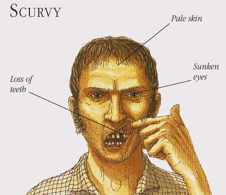
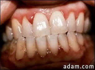
Scurvy leads to the formation of liver spots on the skin, spongy gums, and bleeding from all mucous membranes. The spots are most abundant on the thighs and legs, and a person with the ailment looks pale, feels depressed, and is partially immobilized. In advanced scurvy there are open, suppurating wounds and loss of teeth.
Scurvy was at one time common among sailors, pirates and others who were on ships that were out to sea longer than perishable fruits and vegetables could be stored and by soldiers who were similarly separated from these foods for extended periods. It was described by Hippocrates (c. 460 BC–c. 380 BC). Its cause and cure has been known in many native cultures since prehistory.
For example, in 1536, the French explorer Jacques Cartier, exploring the St. Lawrence River, used the local natives' knowledge to save his men who were dying of scurvy. He boiled the needles of the arbor vitae tree (Eastern White Cedar) to make a tea that was later shown to contain 50 mg of vitamin C per 100 grams. However it was a Scottish surgeon in the British Royal Navy, James Lind (1716–1794) who first proved it could be treated with citrus fruit in experiments he described in his 1753 book, A Treatise of the Scurvy.
In infants, scurvy is sometimes referred to as Barlow's disease, named after Sir Thomas Barlow (1845–1945), a British physician who described it. Barlow's disease is different from Barlow's syndrome.
Scurvy in infants
Scurvy can be prevented by a diet that includes citrus fruits such as limes, oranges, or lemons. Other good sources of Vitamin C are fruits such as guava, papaya, tomatoes or strawberries. It can also be found in some vegetables, such as bell peppers, broccoli, potatoes, cabbage, spinach, paprika, and even pickles. Though redundant in the face of a balanced diet, various nutritional supplements are available that provide ascorbic acid well in excess of that required to prevent scurvy, and even some candies contain vitamin C.
Tuesday, May 22, 2007
Symptoms of Vitamin B Deficiency and Why Vitamin Pills Are Not Enough
Before delving into the problems of vitamin B complex deficiency and its symptoms, it is first important to understand the difference between vitamins that are in pill form versus vitamins that are contained within nature’s foods. There is a world of difference — a difference between sickness and health. Vitamins just do not work like foods, and foods are what our bodies were designed to use for healing, prevention and energy. There is no substitute, and no matter how you look at it, vitamin pills are an invention of scientists, so they are prone to cause side effects, be incomplete and lack what we need to overcome our health problems.
Vitamins have been sold and marketed as the “magic bullet” for all health problems, yet food science researchers are bringing out the truth about vitamins, minerals, multivitamins and antioxidants— and it’s not an easy pill to swallow. The problem is that vitamins, when not still contained in their original food (oranges, bananas, spinach, broccoli, etc.) are merely chemicals. Our bodies do not recognize vitamins as nutrients, because they don’t work the same way as whole foods for these simple reasons:
Foods contain not just vitamins, but the co-workers (synergists) and helper nutrients that allow vitamins to work
Foods are never found in high potency, so you won’t suffer any toxic side effects that have been proven to exist with ALL vitamin pills. As one expert stated, “Foods never deliver toxic doses [of vitamins]. (Hamilton, p.205)
Vitamins are just a small part of what our bodies require for health and healing. It is very often that it is the other food properties that help us while the vitamins are secondary.
Vitamin pills need other nutrients in order to work.
For these reasons, and more, vitamin pills, despite their use and overuse, are not turning people’s health around. They are lacking the properties of real nutrition which can only come from eating nature’s real, whole, raw foods. The only supplement that someone should take, therefore, is a whole food formula WITHOUT any isolated (singular vitamin). This is an important point, because most supplements called “whole food” are combinations of real foods and isolated vitamins. You have to carefully read the labels to see. Look for these words to identify vitamin chemicals on a label:
Pyridoxine
Thiamine or thiamin
Niacin or niacinamide
Palmitate
ascorbic acid
Riboflavin
Mixed tocopherols
B12
cyanocobalamin
more
Vitamins are not foods, so instead of vitamins and minerals on a label, you should be looking for the names of foods and herbs on the label. Don’t be fooled by high milligrams, high potency, standardization or any other such terms that just do not apply to real foods from nature.
VITAMIN B COMPLEX DEFICIENCY:
MAJOR CAUSES
Vitamin B complex comprises a number of vitamins that exist as a family. They should not be taken individually. In this modern era, millions of people suffer from a deficiency of vitamin B for several reasons, chief among which are:
Stress: emotional, physical and spiritual
Processed foods in the diet: these are not real foods and so they tax the body
Refined sugar. The average person consumes at least 140 pounds of sugar a year which robs the body of its vitamin B stores
Drugs: both recreational and prescription drugs deplete vitamin B
Toxins: poisons in the environment and personal care products deplete vitamin B complex
Malnutrition. Most people are malnourished because they are not eating the right kinds of foods
Cooking. Most people do not eat enough real, raw foods, so vitamin B is killed or so depleted that people are not getting enough of it in the diet
DO YOU HAVE ANY OF THESE SYMPTOMS?
Once your body has been deprived of the vitamin B complex due to reasons stated above, it begins to show symptoms (signs) of altered, diminished or poor health. This is because the vitamin B complex (within foods, not vitamin pills) is responsible for such a wide variety of activities, including cellular differentiation, transmission of nerve electricity, health of nerve cells, heart pulse rate, muscular contraction, digestion, brain function, thought processes and energy production. Without adequate vitamin B complex from foods, you can experience one or more of any one of these symptoms:
mental problems
heart palpitations
heart arrythmias
fibrillation
indigestion
chronic fatigue
chronic exhaustion
paranoia, vague fears, fear that something dreadful is about to happen
nervousness
ADD (attention deficiency), inability to concentrate, irritability
feeling of uneasiness
thoughts of dying
easy agitation, frustration
inability to sleep (insomnia)
restlessness
tingling in hands
tingling fingers and toes
rashes
crying spells, inability to cope
soreness all over
and so much more.
CHRONIC VITAMIN B DEFICIENCY
Vitamin B deficiency can sneak up on you, because it doesn’t have to create serious health problems right away. In fact, medical researchers have discovered that very often there can be no detectable signs according to scientific instrumentation, that you are experiencing a deficiency. For example, “memory impairment due to vitamin B12 deficiency can precede blood symptoms of deficiency by years. Evidence that vitamin B12 deficiency accounts for some cognition deficits in older people comes from a study that revealed abnormal short-term memory in more than two-thirds of clients with pernicious anemia…The researchers recommend that a diagnosis of senile dementia should not be made, even in the absence of anemia, until vitamin B12 status is determined biochemically.” (Hamilton, p. 476) This means that it is possible that certain mental disorders can be directly attributable to vitamin B complex deficiency, and it is easier to first start replenishing stores of vitamin B complex than to begin treating difficult mental illnesses with drugs, therapy or psychological counseling.
When vitamin B deficiency becomes chronic (long-lasting), other problems can occur, including troubles with your adrenal system. The adrenal glands serve many purposes, but in relation to this topic, they are the back up system for making energy. When there is a chronic lack of vitamin B complex then the adrenal glands are called upon to produce quick energy by injecting certain hormones like adrenaline into your system so that you can cope with life. If this goes on for a long time, then the adrenal glands become impaired or worn out, leading to even more health problems.
WHAT YOU CAN DO TO HELP YOURSELF
There are a number of things you can do to prevent and overcome vitamin B complex deficiency:
1. Stop taking vitamin pills and switch over to NutriPlex Whole Food Formulas products. The three most recommended to support you are: BFood Complex, AdrenaPlex and SuperGreens PhytoFood. These products are made only of foods and not parts of foods or isolated vitamins. They won’t add to your problems the way vitamin pills can, and they are not toxic the way vitamin pills can be.
2. Stop eating refined sugar. Click here to read all about refined sugar and its bad effects from researcher Nancy Appleton, PhD.
3. Stop eating artificial ingredients. Read all labels on your foods and if there are names of chemicals, don’t eat them. (Read the author’s book, Illness Isn’t Caused by a Drug Deficiency. Switch to organic foods.
4. Reduce stress through a regular exercise program, meditation, counseling and/or hobbies
5. Stop drinking coffee; switch to organic decaffeinated coffee or organic green tea
6. Eat more vitamin B-containing foods — oats, barley, wheat bran, avocado, salmon, Brazil nuts and others.
7. Be patient. It took a while to create a vitamin B deficiency, so it takes a while to reverse the problem; with severe cases it can take a year or so, with milder cases it can take just a few days.
8. If you have a friend or relative with a mental or emotional disorder, think of the possibility of a vitamin B complex deficiency and then make suggestions for adjustments in their diet and take appropriate whole food formulas.
9. Always coordinate all of your health and treatment plans with all of your practitioners.
10. Avoid toxins in your life. This is a serious issue that causes more health problems than people commonly realize. (See the author’s book, Evil Genius in the Garden of Eden — how toxins make us sick and corporations profit from our illness).
Sources:
Journal of the American Medical Association (JAMA); “Vitamins for Chronic Disease Prevention in Adults,” Clinical Applications, Robert H. Fletcher, MD,MSc; Kathleen M. Fairfield, MD,DrPH
JAMA. 2002;287:3127-3129.
Shayne, PhD, Vic, Man Cannot Live on Vitamins Alone, 2004
Shayne, PhD, Vic, Illness Isn’t Caused by a Drug Deficiency!, 2001
Hamilton, et.al, Nutrition Concepts & Controversies, 5th Ed., West Publishing, St. Paul, 1991
Stryer, PhD. Lubert, Biochemistry, 2nd Ed., Stanford University, WH Freeman & Co., San Francisco, 1981
About the AuthorDr. Vic Shayne is a doctor-consultant and food science researcher and author whose website is www.nutritionresearchcenter.org. ©2005 Dr. Vic Shayne. This article may not be used in conjunction with the promotion or sale of any product unless authorized in writing by the author.
Tuesday, May 15, 2007
Rickets
thinking RICKETTS?
well, you're WRONG. It's more like R-I-C-K-E-T-S. this:
and this:
By adding more amounts of vitamin D and calcium to the diet would generally correct any bone problems resulting from rickets. However, hereditary rickets require specialised treatment and skeletal deformaties resulting from rickets may need corrective surgery.
thinking RICKETTS?
well, you're WRONG. It's more like R-I-C-K-E-T-S. this:
and this:
Vitamin D is needed in promoting absorption of calcium and phosphorus, from the gastrointestinal tract, which children need to build strong bones. A deficiency of vitamin D makes it difficult to maintain balanced calcium and phosphorus levels in your bones.
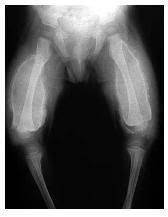
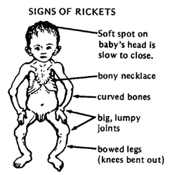
The body senses an imbalance of calcium and phosphorus in the bloodstream and hence reacts by taking calcium and phosphorus from the bones to raise blood levels until they are balanced. This softens and weakens the bone structure, resulting most commonly in skeletal deformities such as bowlegs or improper curvature of the spine.
Causes:
- stay indoors too much, or live in climates with little exposure to sunlight. (vitamin D can be produced by the skin when it is exposed to sunlight)
- malabsorption disorders (they reduce the absorption of fats, which will in turn decrease the ability of vitamin D to be absorbed by the body)
- if you follow a strict vegetarian diet
- lack of calcium and phosphorous in diet
- lactose intolerant (have difficulty digesting milk products)
- breastfed without taking vitamin D supplements (breast milk does not supply the needed amount a vitamin D)
- dark skin
- kidney disorders (renal tubular acidosis)
- can't make or use vitamin D because of a medical disorder such as celiac disease
- through genes (hereditary rickets is passed down through families)
By adding more amounts of vitamin D and calcium to the diet would generally correct any bone problems resulting from rickets. However, hereditary rickets require specialised treatment and skeletal deformaties resulting from rickets may need corrective surgery.
Wednesday, May 9, 2007
Carnitine Deficiency
Carnitine is a naturally occurring hydrophilic amino acid derivative, produced endogenously in the kidneys and liver and derived from meat and dairy products in the diet. It plays an essential role in the transfer of long-chain fatty acids into the mitochondria for beta-oxidation. Carnitine binds acyl residues and helps in their elimination, decreasing the number of acyl residues conjugated with coenzyme A (CoA) and increasing the ratio between free and acylated CoA.
Carnitine deficiency is a metabolic state in which carnitine concentrations in plasma and tissues are less than the levels required for normal function of the organism. Biologic effects of low carnitine levels may not be clinically significant until they reach less than 10-20% of normal. Carnitine deficiency may be primary or secondary.Primary carnitine deficiency is caused by a deficiency in the plasma membrane carnitine transporter, with urinary carnitine wasting causing systemic carnitine depletion. Intracellular carnitine deficiency impairs the entry of long-chain fatty acids into the mitochondrial matrix. Consequently, long-chain fatty acids are not available for beta-oxidation and energy production, and the production of ketone bodies (which are used by the brain) also is impaired.
Regulation of the intramitochondrial free CoA also is affected, with accumulation of acyl-CoA esters in the mitochondria. This, in turn, affects the pathways of intermediary metabolism that require CoA (eg, Krebs cycle, pyruvate oxidation, amino acid metabolism, mitochondrial and peroxisomal beta oxidation).
The 3 areas of involvement include (1) the cardiac muscle, which is affected by progressive cardiomyopathy (by far, the most common form of presentation), (2) the central nervous system, which is affected by encephalopathy caused by hypoketotic hypoglycemia, and (3) the skeletal muscle, which is affected by myopathy.
Muscle carnitine deficiency (restricted to muscle) is characterized by depletion of carnitine levels in muscle with normal serum concentrations. Evidence indicates that the causal factor is a defect in the muscle carnitine transporter.
In secondary carnitine deficiency, which is caused by other metabolic disorders (eg, fatty acid oxidation disorders, organic acidemias), carnitine depletion may be secondary to the formation of acylcarnitine adducts and the inhibition of carnitine transport in renal cells by acylcarnitines.
In disorders of fatty acid oxidation, excessive lipid accumulation occurs in muscle, heart, and liver, with cardiac and skeletal myopathy and hepatomegaly. Long-chain acylcarnitines also are toxic and may have an arrhythmogenic effect, causing sudden cardiac death.
Encephalopathy may be caused by the decreased availability of ketone bodies associated with hypoglycemia. Preterm newborns also may be at risk for developing carnitine deficiency because immature renal tubular function combined with impaired carnitine biosynthesis renders them strictly dependent on exogenous supplies to maintain normal plasma carnitine levels.
Valproic acid may cause an acquired type of secondary carnitine deficiency by directly impairing renal tubular reabsorption of carnitine. The effect on carnitine uptake and the existence of an underlying inborn error involving energy metabolism may be fatal; in other cases, it may primarily affect the muscle, causing weakness.
Osteoporosis could be caused by factors other than declining estrogen levels
New evidence in the April 21, 2006 Cell challenges the long-standing notion that declining estrogen levels alone lead to osteoporosis after women go through menopause. The researchers rather found that high levels of pituitary-derived follicle-stimulating hormone (FSH) cause bone loss in mice. The pituitary is a master gland found at the base of the brain.
What's more, the researchers reported, mice with symptoms of severe estrogen deficiency, lacking either the FSH hormone or its receptor, became resistant to bone loss.
FSH normally triggers egg development and stimulates estrogen production by the ovaries, the researchers explained. As women approach menopause and estrogen levels decline, the pituitary gland responds by releasing more FSH.
Osteoporosis affects nearly 45 million women worldwide with fracture rates that far exceed the combined incidence of breast cancer, stroke, and heart attacks, the researchers said. The disease results from a disruption of the fine balance between bone formation and resorption. After menopause, resorption exceeds new bone formation, leading to a net bone loss that can be slowed by estrogen therapy through mechanisms that have remained somewhat murky, Zaidi said.
However, emerging evidence has begun to cast some doubt on whether estrogen deficiency can fully explain bone loss after menopause, he added. For example, mice without an intact pituitary gland become resistant to the effects of ovary loss on bone density. FSH levels also show a closer correlation than estrogen levels to the rate of bone turnover in postmenopausal women.
The researchers now show that mice lacking FSH or its receptor become resistant to bone loss despite severe loss of ovarian function. In mice with normal ovaries and approximately half the normal concentration of FSH, bone mass increased due to a decline in bone resorption by cells known as osteoclasts, which break down bone. Indeed, they report, FSH stimulates receptors found on the surface of bone-degrading osteoclasts and their precursors, leading to the bone cells' formation and function.
Saturday, April 21, 2007
Biotinidase Deficiency
Biotinidase deficiency is an inherited disorder in which the body is not able to process the vitamin biotin properly. Biotin is an important water-soluble vitamin that aids in the metabolism of fats, carbohydrates and proteins. 1 in every 110, 000 babies will have the deficiency.
Newborns suffering from biotinidase deficiency will appear normal at birth. The deficiency develops over time ranging from a few weeks to several years of age.
Biotinidase deficiency is an autosomal recessive disorder affecting both males and females. In individuals with this disorder, both copies of the biotinidase gene are defective. Both parents of an affected child have one abnormal copy of the gene, but usually do not show symptoms because they also have one normal copy. The normal copy provides approximately 50% of the usual enzyme activity, a level adequate for the body's needs. Individuals with one abnormal copy of the gene and 50% enzyme activity are said to be carriers or heterozygotes. As is typical of autosomal recessive inheritance, their risk for having another child with the disorder is 25% in each subsequent pregnancy.
Biotin deficiency can result in behavioural disorders, lack of coordination, learning disabilities, hearing loss, developmental delay and seizure.
Without biotinidase activity, biotin cannot be separated from foods and hence cannot be used by the body. Biotinidase enzyme also recycles biotin from enzymes that are important in metabolism.
HOWEVER, there is no need for much worry as it can be treated with oral biotin supplementation, preventing development of critical symptoms.
Here is the chemical structure of biotin:
Iodine Deficiency
Iodine is an essential trace element (a chemical element that is needed in small quantities for proper growth, development and physiology aka micronutrient). When there is little iodine in our diet, iodine deficiency gives rise to goiter (swelling in the neck - below) and cretinism (severely stunted physical and mental growth) resulting in developmental delays and health problems.
Mild iodine deficiency has been reported to reduce intelligence quotients (I.Q.) by 10%-15% and cause increased rates of stillbirths and infant mortality
So where does the iodine in our diet come from?
Although the element is quite rare, certain plants have the ability to concentrate it from seawater, thus introducing iodine into the food chain.
Iodine is widely available in:
In areas where little or no marine food is eaten, the people will suffer from iodine deficiency. To combat this, iodine deficiency is combatted by the additions of iodine to small amounts of table salt, known as iodized salt, and other foodstuffs such as flour.
Did you know?
Approximately two billion people are affected by iodine deficiency in this world, particularly in India, with 500 million suffering from deficiency.
Here are the recommended intakes for iodine:
Mild iodine deficiency has been reported to reduce intelligence quotients (I.Q.) by 10%-15% and cause increased rates of stillbirths and infant mortality
So where does the iodine in our diet come from?
Although the element is quite rare, certain plants have the ability to concentrate it from seawater, thus introducing iodine into the food chain.
Iodine is widely available in:
- Cod
- Sea bass
- Haddock
- Perch
- Kelp
- Dairy products
- Plants grown in iodine-rich soil
In areas where little or no marine food is eaten, the people will suffer from iodine deficiency. To combat this, iodine deficiency is combatted by the additions of iodine to small amounts of table salt, known as iodized salt, and other foodstuffs such as flour.
Did you know?
Approximately two billion people are affected by iodine deficiency in this world, particularly in India, with 500 million suffering from deficiency.
Here are the recommended intakes for iodine:
- Infants - 40 to 50 micrograms
- Children (1 to 3 YO) - 60 micrograms
- Children (4 to 6 YO) - 90 micrograms
- Children (7 to 10 YO) - 120 micrograms
- Children (11 years and above) - 150 micrograms
- Pregnant women - 175 micrograms
- Adult men and women - 100 to 200 micrograms
Monday, April 16, 2007
Primary Immune Deficiency Disorder
A Primary Immune Deficiency Disorder is present when a body part is missing or is unable to function normally, these are caused by genetic and intermal problems, increasing susceptibility to infections. It is a broad heading for many World Health Organisation recognised deficiencies.
It has been estimated that there are 100 different primary immunodeficiency diseases. All are genetic conditions in which specific cells of the immune system do not function properly. Clinical symptoms range from mild or nonexistent as in the case of selective IgA deficiency to severe symptoms as in the case of severe combined immunodeficiency (SCID), commonly referred to as "bubble-boy" syndrome.
Possible types of Primary Immune Deficiency Disorder:
Antibody Deficiencies An antibody is a protein made by white blood cells called B lymphocytes and plasma cells. Its function is to recognize and mark foreign microorganisms. It can be diagnosed with frequent and severe infections caused by organisms that do not affect people with healthy immune systems.
Complement Deficiencies The complement system consists of a group of proteins that attach to bacteria and viruses.
These proteins work together to disable and kill invading pathogens. People deficient in one or another of these proteins have a diminished ability to kill invaders, resulting in increased infections. Such people may also develop antibodies that react against the body's own cells and tissues, resulting in autoimmune diseases with immune-mediated damage to the body. The most common of these deficiencies is:
C2 Deficiency-an inherited defect in the gene for the complement protein called C2. This defect can cause an autoimmune disease such as systemic lupus erythematosus or can result in severe infections such as meningitis. The illnesses usually appear in childhood or in early adulthood.
Phagocytic Cell Deficiencies Phagocytes: white blood cells called neutrophils and macrophages that engulf and kill microorganisms. If defective either in their ability to kill pathogens or in their ability to move to the site of an infection. An increase in infections can happen. The most severe form is:
Chronic Granulomatous Disease-inherited deficiencies of molecules needed by neutrophils to kill certain infectious organisms. These illnesses usually appear early in childhood but may appear as late as adolescence. People with chronic granulomatous disease develop frequent and severe infections of the skin, lungs, and bones and develop localized, swollen collections of inflamed tissue called granulomas.
Due to healthcare advances, most patients with a Primary Immune Deficiency Disorder will be able to live normal lives to adulthood; however, they will have to rely on life-long treatment, such as intravenous gamma globulin infusions, antibiotic therapies, or bone marrow transplantation. Although the susceptibility to infections is a major consequence of the primary immunodeficiency diseases, they may cause other health problems as well, including allergies, asthma, swollen joints, digestive tract problems, growth problems or an enlarged liver and spleen.
The primary immune deficiency disorders range in frequency. Some disorders such as selective IgA deficiency are quite common, occurring as often as 1/500 to 1/1000 individuals. Others disorders such as SCID are rare affecting perhaps one person per million. Approximately 25,000 to 50,000 Americans are severely affected by primary immune deficiency disorders.
For more information:
http://www.primaryimmune.org/pid/1996_who.pdf -a very difficult report from the WHO.
Source:
http://www.primaryimmune.org/pid/whatis_pid.htm
http://www.medhelp.org/NIHlib/GF-739.html
http://www.medterms.com/script/main/art.asp?articlekey=25189
http://www.aaaai.org/patients/topicofthemonth/0407/
Sunday, April 15, 2007
Arginase Deficiency
Arginase deficiency is thought to be the least common of the urea cycle disorders. Two separate isozymes of the enzyme arginase exist. Type I is found in the liver and contributes the vast majority of hepatic arginase activity, while type II is inducible and found in extrahepatic tissues. The disease is caused by deficiency of arginase type I in the liver.
The hepatic urea cycle is the major route for waste nitrogen disposal, which is generated chiefly from protein and amino acid metabolism. Low-level synthesis of certain cycle intermediates in extrahepatic tissues makes a small contribution to waste nitrogen disposal as well. A portion of the cycle takes place in mitochondria. Overall, activity of the cycle is regulated by the rate of synthesis of N-acetylglutamate, the enzyme activator that initiates incorporation of ammonia into the cycle.
The reaction normally mediated by arginase is the terminal step in the urea cycle, which liberates urea with regeneration of ornithine. Consequently, as in argininosuccinic aciduria, both waste nitrogen molecules normally eliminated by the urea cycle are incorporated into the arginine substrate molecule in the reaction.
Think that is complicated? Look at THIS.
As an inherited disorder, the age of onset is typically during the neonatal period(or in simpler terms, the early part of infancy). Because of its atypical form of manifestation, the disease may be easily missed in the neonatal period and only recognized in later infancy or early childhood. Some cases likely go undiagnosed, with clinical symptomatology attributed to cerebral palsy. Also, being an inherited disorder, parents have 1 in 4 chances of having an affected child with each conception. Affected infants and children may exhibit mental retardation, seizures, and spasticity (difficulty in walking).Luckily, arginase deficiency hardly results in death. yay!
Diagnosis is made by special blood tests called plasma amino acids. Enzyme level can be measured in the blood however specialized laboratory help is required for this. In some situations DNA mutation analysis is also available. To prevent giving birth to an affected child, prenatal diagnosis is possible based on DNA mutation analysis.
But if YOU have arginase deficiency (touch wood), this is what you should do!
1. Normalize plasma arginine levels
2· Go for genetic counselling
3· Take a low protein diet
4· Take these drugs: Sodium phenyl butyrate/ benzoate to divert nitrogen from urea cycle
Sunday, April 1, 2007
Introduction
A deficiency disease is basically a disease, such as rickets or scurvy, that is caused by a dietary deficiency of specific nutrients, especially a vitamin or mineral. The disease may stem from insufficient intake, digestion, absorption, or utilization of a nutrient.
In our following posts, we shall elaborate on several defiency diseases known to man.
Subscribe to:
Posts (Atom)